- This topic is empty.
-
AuthorPosts
-
2026-11-12 at 7:32 pm #85068
■ 1. Background
H-151 (T5674), CAS: 941987-60-6 is a highly potent and selective antagonist of the stimulator of interferon genes (STING) adaptor that acts through covalent chemistry at a critical regulatory residue on the protein. In the canonical cytosolic DNA–sensing axis, DNA engagement activates cyclic GMP–AMP synthase (cGAS), which generates 2′3′-cGAMP. cGAMP then binds ER-resident STING, triggering its conformational change, exit from the ER, and trafficking to the Golgi, where STING becomes palmitoylated and organizes signaling complexes that activate TANK-binding kinase 1 (TBK1), leading to IRF3 phosphorylation and type I interferon gene induction, with parallel activation of NF-κB–dependent inflammatory programs [1,2]. A decisive biochemical checkpoint in this cascade is STING palmitoylation at Cys91 on the cytosolic face of the Golgi.
H-151 directly targets this checkpoint. It covalently binds Cys91 of STING and thereby prevents the palmitoylation of the same residue, a lipid modification required for productive assembly of downstream signaling scaffolds. By locking Cys91 in a non-palmitoylatable state, H-151 functionally disengages STING from TBK1 and IRF3 activation, suppressing the phosphorylation events and transcriptional outputs (type I interferons and interferon-stimulated genes, as well as NF-κB–driven cytokines) that normally follow cGAMP-dependent STING engagement. Because the intervention occurs at the shared palmitoylation node, H-151 can blunt STING signaling elicited by diverse upstream stimuli that converge on STING, offering a pathway-level blockade independent of the specific DNA source or cGAMP origin.
This mechanism affords several research utilities. First, H-151 provides a temporal, chemically precise means to acutely inhibit STING in cells and organisms, enabling dissection of STING dependence for innate responses to cytosolic DNA, mitochondrial genome leakage, or genotoxic stress. Second, by acting on Cys91, H-151 is suited to mechanistic studies of post-translational control of STING, for example by correlating loss of palmitoylation with diminished TBK1 recruitment, reduced IRF3 nuclear translocation, and attenuated interferon-stimulated gene signatures. Third, in vivo and ex vivo use of H-151 facilitates modeling of interferon-driven autoinflammatory processes in which aberrant STING activation is implicated, allowing researchers to calibrate phenotypes and transcriptional programs to the degree and timing of STING engagement. Typical experimental readouts include STING palmitoylation status, TBK1/IRF3 phosphorylation, IFNB1 and ISG transcript abundance, secreted interferon levels, and NF-κB–responsive cytokines, alongside orthogonal controls such as cGAS perturbation to position the H-151 effect within the cGAS–STING axis.
Altogether, H-151 exemplifies a pathway-informed chemical probe that disables STING by covalently occupying Cys91 to block its palmitoylation, thereby silencing the core signaling events that link cytosolic DNA sensing to interferon and inflammatory gene expression. Its selectivity and defined molecular point of action make it a valuable tool for delineating STING’s roles across innate immune biology and for interrogating autoinflammatory mechanisms in vivo and ex vivo [1,2].
■ 2. Literature review
■ 2.1 Targeted Inhibition of cGAS/STING signaling induced by aberrant R-Loops in the nucleus pulposus to alleviate cellular senescence and intervertebral disc degeneration
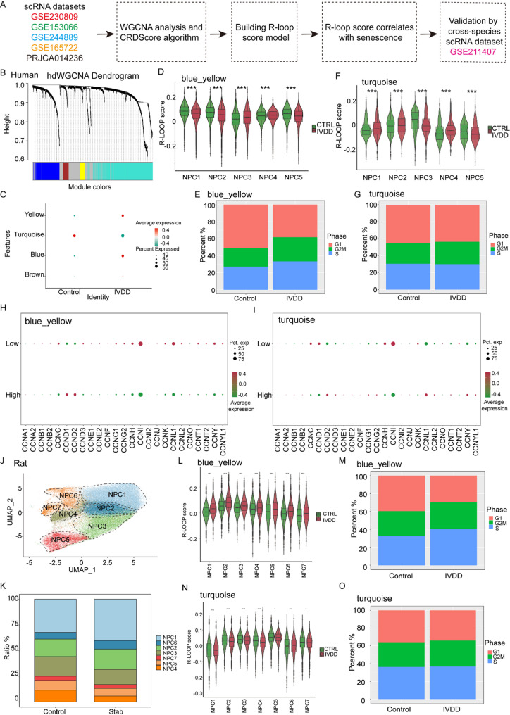
▲ (A) Illustration of establishing the R-Loop score model based on scRNA sequencing; (B) Weighted gene co-expression network analysis (WGCNA) dendrogram of 1,185 R-Loop regulators (deeper colors indicate stronger interactions); (C) Correlation analysis of the four modules generated from WGCNA analysis in relation to IVDD; (D) Differences in the turquoise module R-Loop score for each NPC cluster between the control and IVDD groups; (E) Cell cycle analysis of NPCs based on the R-Loop score for the turquoise module; (F) Differences in the yellow-blue R-Loop score for each NPC cluster between the control and IVDD groups; (G) Cell cycle analysis of NPCs based on the R-Loop score for the yellow-blue module; (H) Differences in cyclin gene expression between the two groups for the turquoise module; (I) Differences in cyclin gene expression between the two groups for the yellow-blue module; (J) UMAP visualization of rat IVD cells as seven NPC subclusters (each dot represents a single cell, colored by subcluster); (K) Relative proportion of each NPC cluster between the control and stab groups (IVDD); (L) Differences in yellow-blue module R-Loop score for each NPC cluster between the control and stab groups; (M) Cell cycle analysis of NPCs based on the R-Loop score for the yellow-blue module; (N) Differences in turquoise R-Loop score for each NPC cluster between the control and stab groups; (O) Cell cycle analysis of NPCs based on the R-Loop score for the turquoise module. NPC: Nucleus pulposus; IVDD: Intervertebral disc degeneration.
H-151 (T5674) was used in this study as a selective small molecule STING antagonist at 1 µM following ERCC5 overexpression in nucleus pulposus cells, as described by the authors. In this setup, immunofluorescence analysis provided direct experimental confirmation that, under STING-inhibited conditions established with H-151, the ratio of senescent nucleus pulposus cells induced by ERCC5 overexpression was reduced. These findings position H-151 within the workflow as the agent implementing STING inhibition at a defined concentration and link this intervention to a measurable decrease in senescence ratios assessed by immunofluorescence staining in the ERCC5 overexpression context.[3]
■ 2.2 Lovastatin-Induced Mitochondrial Oxidative Stress Leads to the Release of mtDNA to Promote Apoptosis by Activating cGAS-STING Pathway in Human Colorectal Cancer Cells
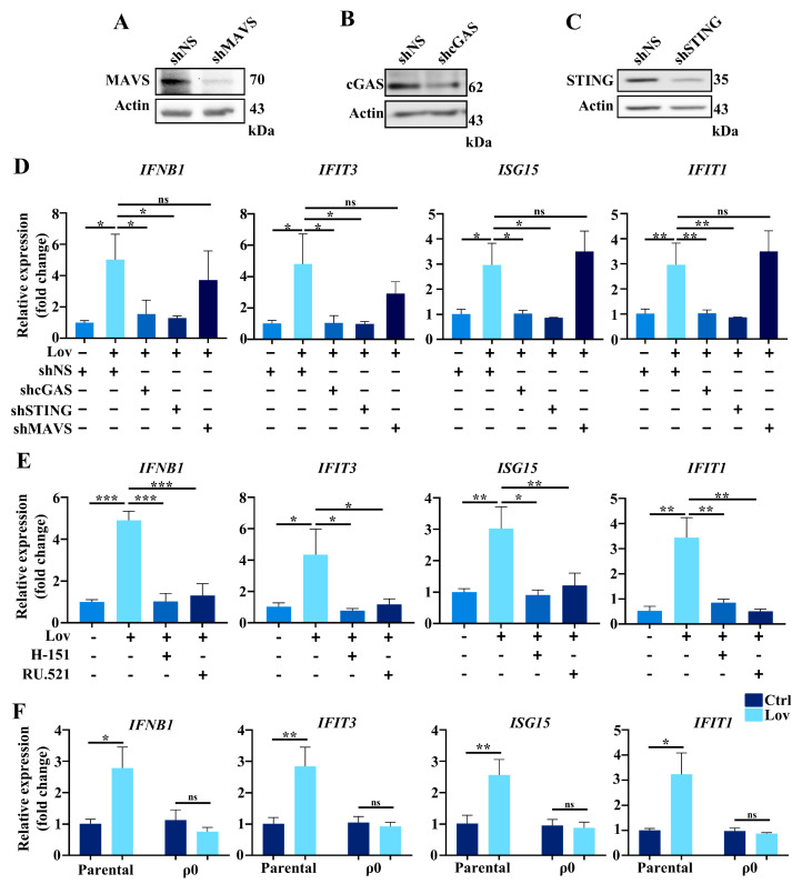
▲ (A–C) Western blotting analysis of MAVS (A), cGAS (B), and STING (C) after lentiviral knockdown in HCT116 cells. β-Actin served as a loading control. (D) The expressions of IFNB1, IFIT3, ISG15, and IFIT1 were quantified by qPCR in cGAS, STING, and MAVS knockdown cells treated with 10 μM lovastatin for 48 h. (E) HCT116 cells were pretreated either with STING inhibitor H-151 (1.5 μM) or cGAS inhibitor RU.521 (1.0 μM) for 2 h, and afterward treated with lovastatin for 24 h; the expressions of IFNB1, IFIT3, ISG15, and IFIT1 were quantified by qPCR. (F) The expressions of IFNB1, IFIT3, ISG15, and IFIT1 were quantified by qPCR in parental and ρ0 HCT116 cells treated with 10 μM lovastatin for 48 h. All data were obtained from at least three independent experiments; * p < 0.05; ** p < 0.01; *** p < 0.001; ns, no significance. The results demonstrate that knockdown of cGAS/STING or treatment with H-151 significantly suppresses lovastatin-induced expression of interferon-stimulated genes, whereas knockdown of MAVS does not, indicating that lovastatin-induced type I IFN activation relies on the cGAS/STING axis rather than MAVS.
H-151 (T5674) was used in this study as a STING inhibitor to interrogate lovastatin-induced signaling in human colorectal cancer HCT116 cells. The investigators blocked the pathway with H-151 and evaluated type I interferon gene responses, stating that these inhibitors prevented the expression of IFNB1, IFIT3, ISG15, and IFIT1 in response to lovastatin. The experimental setup specified that HCT116 cells were pretreated with H-151 at 1.5 μM for 2 hours, then exposed to lovastatin for 24 hours, and transcript levels of IFNB1, IFIT3, ISG15, and IFIT1 were measured by qPCR. Under these conditions, H-151 prevented the induction of these interferon-related genes by lovastatin. Together, the data identify H-151 as a STING inhibitor within this system and show that its application blocks lovastatin-driven type I interferon gene expression in HCT116 cells using a defined pretreatment regimen and qPCR-based readouts.[4]
■ 2.3 A Single‐Atom Manganese Nanozyme Mn‐N/C Promotes Anti‐Tumor Immune Response via Eliciting Type I Interferon Signaling
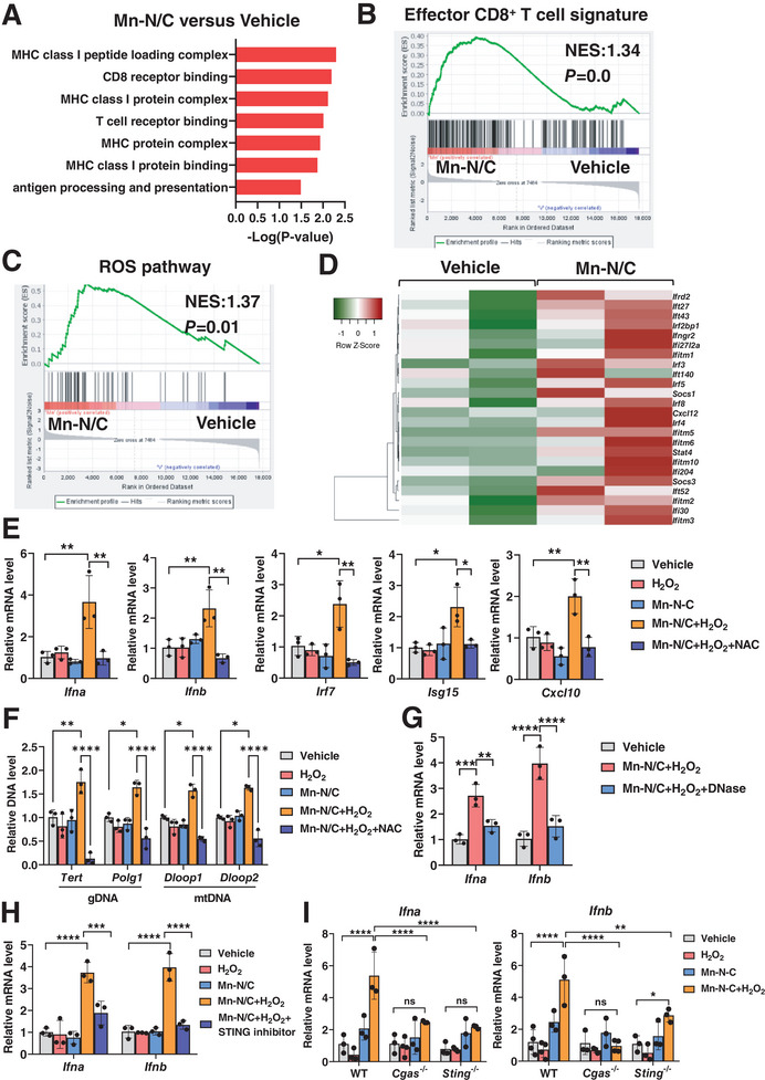
▲ Mn-N/C activates type I IFN signaling by cGAS/STING-dependent cytosolic DNA sensing. (A) GO enrichment gene analysis of pathways in tumor tissues of MC38-bearing mice with Vehicle or Mn-N/C treatment. (B) GSEA analysis of enrichment of CD8+ T cell effector signatures in tumor tissues of MC38 tumor-bearing mice with Vehicle or Mn-N/C treatment. (C) GSEA analysis of enrichment of ROS pathway signatures in tumor tissues of MC38-bearing mice with Vehicle or Mn-N/C treatment. (D) Heatmap of type I IFN gene signatures in tumor tissues of MC38-bearing mice with Vehicle or Mn-N/C treatment. (E) Real-time PCR analysis of Ifna, Ifnb, Irf7, Isg15, and Cxcl10 gene expression levels in MC38 tumor cells treated with H₂O₂, Mn-N/C, or plus NAC (1 mM) for 24 h. (F) Cytosolic amount of gDNA and mtDNA in MC38 tumor cells treated with H₂O₂, Mn-N/C, or plus NAC (1 mM) for 24 h. (G) Real-time PCR analysis of Ifna and Ifnb expression in MC38 tumor cells treated with Mn-N/C and H₂O₂, or plus DNase for 24 h. (H) Real-time PCR analysis of Ifna and Ifnb expression in MC38 tumor cells treated with Mn-N/C, H₂O₂, or plus STING inhibitor H-151 for 24 h. (I) Real-time PCR analysis of Ifna and Ifnb expression in WT, Cgas⁻/⁻, Sting⁻/⁻ MC38 tumor cells treated with H₂O₂ or Mn-N/C for 24 h. The results demonstrate that Mn-N/C-induced type I IFN signaling is dependent on the cGAS/STING pathway, and H-151 significantly blocks this signaling.
H-151 (T5674) was used as a STING inhibitor to probe the role of cGAS–STING signaling in type I interferon responses triggered under oxidative conditions. In MC38 tumor cells exposed to Mn‑N/C and H2O2, pre-treatment with H-151 led to a significant suppression of type I interferon induction, as determined by gene expression readouts. This effect was specifically observed when cells were subjected to Mn‑N/C plus H2O2 and then evaluated, indicating that H-151 effectively countered the upregulation of type I interferon signaling in this context. The study quantified Ifna and Ifnb expression by real-time PCR after 24 hours of exposure, explicitly including conditions with H-151 alongside Mn‑N/C and H2O2, which established the time frame and assay parameters for detecting the impact on interferon gene expression. Experimentally, H-151 was applied at 1 µM together with Mn‑N/C and H2O2 in vitro, further specifying the conditions under which the suppression of type I interferon responses occurred. Collectively, these findings show that H-151 functioned as a STING inhibitor in this system and significantly reduced the Mn‑N/C plus H2O2–induced elevation of Ifna/Ifnb transcripts in MC38 tumor cells under the reported assay conditions.[5]
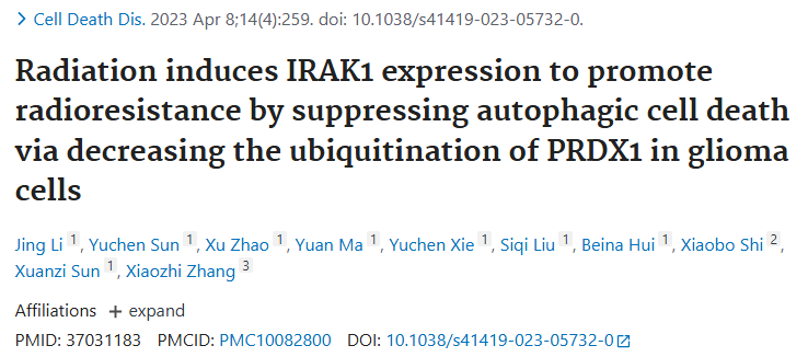
■ 2.4 Radiation induces IRAK1 expression to promote radioresistance by suppressing autophagic cell death via decreasing the ubiquitination of PRDX1 in glioma cells
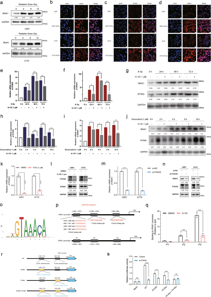
▲ cGAS-STING signaling participates in radiation-induced IRAK1 expression. (a) U251 and A172 glioma cells were exposed to a range of X-ray IR doses. IRAK1 protein expression levels were detected by Western blotting at 48 h post-IR exposure. (b–d) Immunofluorescence staining was performed to determine the expression abundance of cGAS (b), STING (c), and IRAK1 (d) in response to 8 Gy IR (scale bar = 25 μm). (e, f) qRT-PCR measurement of STING (e) and IRAK1 (f) mRNA expression in U251 cells treated with 8 Gy X-ray irradiation ± H-151 (1 μM). (g) The levels of IRAK1 and STING were assessed by Western blotting in U251 cells at 0, 24, 48, and 72 h after 8 Gy irradiation ± H-151 (1 μM). (h, i) qRT-PCR measurement of STING (h) and IRAK1 (i) mRNA expression in U251 cells treated with doxorubicin (1 μM) ± H-151 (1 μM). (j) Effects of doxorubicin (1 μM) ± H-151 (1 μM) treatment on IRAK1 and STING protein expression were evaluated by Western blotting in U251 cells. (k, l) The influence of H-151 (1 μM) on FOXA2 was measured by RT-qPCR (k) and Western blotting (l). (m, n) Changes in the mRNA (m) and protein (n) levels of IRAK1 caused by FOXA2 knockdown in glioma cells. The gray value ratios of the corresponding proteins/GAPDH are shown under the lanes. (o, p) The FOXA2-binding motif was predicted by JASPAR, and schematic images of the potential FOXA2 binding sites in the IRAK1 promoter region are shown. (q) ChIP-qPCR analysis of FOXA2 binding on the IRAK1 promoter in U251 cells. (r, s) Luciferase reporter assay for detecting the activity of wild-type or mutant IRAK1 promoters in U251 cells transfected with FOXA2 siRNA or Scramble siRNA. Data are shown as mean ± SD from three independent experiments; *p < 0.05, **p < 0.01, ***p < 0.001.
H-151 (T5674) was utilized as a STING inhibitor in this study. Cellular inhibitory activities of H-151 on STING after 8 Gy irradiation were verified by RT-qPCR and Western blotting in U251 and A172 glioma cells. Ionizing radiation markedly induced IRAK1 expression, which was decreased by H-151. Consistently, doxorubicin-induced IRAK1 expression was remarkably reduced by H-151 in U251 and A172 cells. Beyond its effects on IRAK1, H-151 significantly suppressed the mRNA and protein levels of the transcription factor FOXA2 in glioma cells, as shown by RT-qPCR and Western blotting. In line with these transcriptional effects, the binding of FOXA2 to the P2 and P3 sites within the IRAK1 promoter region was impaired following H-151 exposure, indicating that H-151 diminished FOXA2 engagement at these regulatory motifs. Together, these experimental findings demonstrate that H-151 functionally inhibits STING signaling in irradiated glioma cells, lowers stress-induced IRAK1 expression under both ionizing radiation and doxorubicin exposure, reduces FOXA2 abundance, and interferes with FOXA2 occupancy at key promoter elements of the IRAK1 gene.[6]
■ 3. Conclusion
H-151 is a highly selective STING antagonist that covalently binds to the Cys91 residue of STING, thereby preventing its essential palmitoylation. This modification blockade disrupts the assembly of downstream signaling complexes, leading to inhibition of TBK1 activation, IRF3 phosphorylation, and the resulting transcriptional induction of type I interferons and NF-κB–dependent inflammatory cytokines. Literature reports demonstrate H-151’s efficacy in various cellular models, including suppression of senescence in nucleus pulposus cells, inhibition of lovastatin-induced interferon responses in colorectal cancer cells, reduction of oxidative stress–triggered interferon signaling in tumor cells, and attenuation of radiation- and chemotherapy-induced stress signaling in glioma cells. These findings establish H-151 as a powerful chemical probe to dissect STING-dependent innate immune pathways and autoinflammatory mechanisms. Future research may explore its utility in modulating pathological inflammation in vivo, refining therapeutic strategies targeting STING-driven diseases, and further elucidating post-translational regulation of STING signaling.
■ 4. Q&A
Q1: What is the molecular target and mechanism of action of H-151?
A1: H-151 covalently binds to the Cys91 residue of STING, preventing its palmitoylation which is essential for downstream signaling. This blockade inhibits TBK1 activation, IRF3 phosphorylation, and the transcription of type I interferons and NF-κB–dependent cytokines.
Q2: How has H-151 been utilized in experimental studies according to the literature?
A2: H-151 has been applied as a selective STING inhibitor in various models: reducing senescence in nucleus pulposus cells following ERCC5 overexpression; blocking lovastatin-induced interferon gene expression in colorectal cancer cells; suppressing oxidative stress–induced interferon signaling in MC38 tumor cells; and inhibiting radiation- and chemotherapy-induced IRAK1 and FOXA2 expression in glioma cells.
Q3: What are potential future research directions for H-151 mentioned in the text?
A3: Future directions include using H-151 to modulate pathological autoinflammatory and interferon-driven diseases in vivo, to dissect STING post-translational modifications, and to develop therapeutic strategies targeting STING signaling pathways.
■ 5. Reference
[1] Ishikawa H, Barber GN, et al. STING is an endoplasmic reticulum adaptor that facilitates innate immune signaling. Nature. 2008;455(7213):674-8.
[2] Sun L, Wu J, Du F, Chen X, Chen ZJ, et al. Cyclic GMP-AMP synthase is a cytosolic DNA sensor that activates the type I interferon pathway. Science. 2013;339(6121):786-91.
[3] Wu D, Yan C, Han L, Li F, Xu X, Sun J, et al.. Targeted Inhibition of cGAS/STING signaling induced by aberrant R-Loops in the nucleus pulposus to alleviate cellular senescence and intervertebral disc degeneration. Journal of Nanobiotechnology. 2025;23(1):.
[4] Huang X, Liang N, Zhang F, Lin W, Ma W. Lovastatin-Induced Mitochondrial Oxidative Stress Leads to the Release of mtDNA to Promote Apoptosis by Activating cGAS-STING Pathway in Human Colorectal Cancer Cells. Antioxidants. 2024;13(6):679.
[5] Qiao W, Chen J, Zhou H, Hu C, Dalangood S, Li H, et al.. A Single‐Atom Manganese Nanozyme Mn‐N/C Promotes Anti‐Tumor Immune Response via Eliciting Type I Interferon Signaling. Advanced Science. 2024;11(14):.
[6] Li J, Sun Y, Zhao X, Ma Y, Xie Y, Liu S, et al.. Radiation induces IRAK1 expression to promote radioresistance by suppressing autophagic cell death via decreasing the ubiquitination of PRDX1 in glioma cells. Cell Death & Disease. 2023;14(4):.
http://www.targetmol.com
TargetMol Chemicals Inc. -
AuthorPosts

- You must be logged in to reply to this topic.